
Pakistan Journal of Neurological Pakistan Journal of Neurological
Sciences (PJNS) Sciences (PJNS)
Volume 15 Issue 2 Article 9
6-2020
Movement disorder associated with celiac disease and thyroid Movement disorder associated with celiac disease and thyroid
dysfunction:case report dysfunction:case report
Ramla Nayaib Hashmi
Neurologist,Rabia Moon Institute Of Neurosciences, Karachi. 2.
Ummul Kiram
Saifee and Mamji Hospital , Karachi
Rabbia Tariq
,The Indus Hospital , Karachi
Follow this and additional works at: https://ecommons.aku.edu/pjns
Part of the Neurology Commons
Recommended Citation Recommended Citation
Hashmi, Ramla Nayaib; Kiram, Ummul; and Tariq, Rabbia (2020) "Movement disorder associated with
celiac disease and thyroid dysfunction:case report,"
Pakistan Journal of Neurological Sciences (PJNS)
:
Vol. 15 : Iss. 2 , Article 9.
Available at: https://ecommons.aku.edu/pjns/vol15/iss2/9

MOVEMENT DISORDER ASSOCIATED WITH CELIAC
DISEASE AND THYROID DYSFUNCTION:CASE REPORT
PAKISTAN JOURNAL OF NEUROLOGICAL SCIENCES
30
VOL. 15 (2) APRIL-JUNE 2020
ABSTRACT:
Autoimmune movement disorders are uncommon yet likely treatable conditions. It is challenging to diagnose and
manage these disorders, ultimately leading to a significant morbidity in terms of functional disability of patient. It is
troublesome for practicing neurologists to identify whether the antibodies which are detected during the work up of
these autoimmune movement disorders are merely incidental or have pathogenic role. This will help them to decide
regarding whether to start immunotherapy or not. Here we have discussed two patients of movement disorder who
were found to have positive work up for other autoimmune diseases. Available Literature on autoimmune movement
disorder is very limited and further research on large scale is needed.
KEY WORDS: Autoimmune movement disorder, autoimmune dystonia, gluten ataxia, celiac disease, thyroid
dysfunction
BACKGROUND: Autoimmune movement disorders comprises of a widespread group of neurologic disorders which
occur either in isolation or along with more diffuse autoimmune illnesses. The exact incidence and prevalence of
autoimmune movement disorders is not known as it’s a rare disorder and very limited data is available on it comprising
of few case reports or small sample studies. One estimates suggest that all autoimmune diseases combined affect
about 3% of the US population, thus approximately 10 million people.
1
Many movement disorders occur due
pathologic alterations in the basal ganglia or their connections including caudate, putamen, and pallidum ,
subthalamic nucleus, substantia nigra, pedunculopontine nucleus, although some exceptions can be present. Due to
the fact that pure, isolated autoimmune movement disorders are uncommon, work up for other aetiologies is strongly
recommended.
2
Any movement disorder phenomenology can occue in these disorders and many of the presentations
can mimic neurodegenerative disorders, such as Huntington disease. Disorders may be ataxic, hypokinetic
(Parkinsonism), or hyperkinetic (myoclonus, chorea, and other dyskinetic disorders
3,4
.
Movement disorders are commonly detected in autoimmune diseases. Chorea may present in patients with
anti-NMDAR encephalitis, Sydenham disease, lupus and anti-phospholipid syndrome. Dystonic seizures and
neuromyotonia may precede autoimmune encephalitis. Anti-glycine antibodies are encountered in the pathogenesis of
progressive encephalomyelitis presenting with rigidity and myoclonus. Hashimoto's encephalitis and
opsoclonus-myoclonus syndrome have commonly tremor and myoclonus. Few case reports and review articles also
highlight the association of Celiac disease and autoimmune thyroid disorders with autoimmune movement disorders
5
6.
Celiac disease (CD) approximately accounts for up to 10% of neurological manifestations irrespective to the
presence of malabsorption symptoms
7
According to an article published in 2018, in patients with autoimmune
disorders affecting the central and peripheral nervous system, movement disorder are frequent. They may be found in
autoimmune disorders incited by an infectious agent, like streptococcus in Sydenham's chorea, or in basal ganglia
encephalitis with antibodies against the dopamine-D2 receptors.In these scenarios, chorea or dystonia are usually the
most prominent.In diffuse or limbic encephalitis with antibodies directed against neuronal cell-surface antigens ,
patients also have MD. Anti-NMDA receptor encephalitis is one of the most common and may present with a variety
of MDs, including: chorea, stereotypies, dystonia and myorhythmia while in stiff-person syndrome or cerebellar ataxia
, intracellular enzyme glutamic acid decarboxylase are found most commonly.
8
Clues of when to suspect an
autoimmune etiology are Female patient , Subacute onset, variable course of disease , neurological disease with
multiple foci , autoimmunity personal or family history , Patient having history of cancer or suspicion of new cancer
,Abnormal laboratory supportive tests, i.e., Cerebrospinal fluid , imaging and the response to immunotherapy.
2
A rapid
diagnosis of an autoimmune disorder is utmost as early therapeutic intervention improves long-term prognosis and
Correspondence to: Ramla Nayaib Hashmi Email:[email protected]
Date of submission: February 17, 2020 Date of revision: March 28, 2020 Date of acceptance: April 09, 2020
Dr. Ramla Nayaib Hashmi
1
, Dr. Ummul Kiram
2
, Dr.Rabbia Tariq
3
.
1.
Consultant Neurologist,Rabia Moon Institute Of Neurosciences, Karachi.
2.
Consultant Neurologist,Saifee and Mamji Hospital , Karachi.
3.
Post graduate (MCPS) Trainee,Family Medicine Department ,The Indus Hospital , Karachi.
CASE REPORT

may be life-saving. Treatment usually comprises of some form of immunotherapy and symptomatic therapy of the
abnormal movements with dopamine depleters, dopamine receptor antagonists, or GABAergic drugs. Early-initiated
immunotherapy leads to the best possible outcomes. Moreover, depending on the antibody profile, a search for cancer
may be necessary followed by removal of an underlying tumour for optimal outcome.
3,4
It is challenging to diagnose
such autoimmune disorders due to the delayed clinical suspicion, improper approach to get the relevant systematic
work up and lack of facilities for specific antibodies detection or in paraneoplastic cases getting extensive work up for
occult neoplasm. The whole work up is expensive and time consuming ending mostly in lost to follow up of patients.
For instance, in literature, work up suggested for autoimmune movement disorder include CRMP-5, ANNA-1 (Hu),
ANNA-2 (Ri), GAD65, VGCC antibodies for chorea, ANNA-2 (Ri), CRMP-5, NMDA-R, GAD65, DPPX antibodies for
myoclonus, NMDA-R for dyskinesia ,LGI1, GAD65 for Parkinsonism,GAD65, DPPX, Amphiphysin for Stiff-Person
Spectrum Disorders,ANNA-1 (Hu), PCA-1 (Yo), GAD65, VGCC, CASPR2, mGluR1, DPPX for ataxia
3,4
Despite all these
limitations in diagnosis, early management is very important.
CASE 1:
26 years old female patient with no known previous comorbid, married, mother of 2 children up and about till 2 years
back. She had history of fever 2 years back which was intermittent, high grade for which she was treated empirically.
Fever subsided in few days but she started complaining of difficulty in speech, tremors of neck and upper limbs along
with paroxysmal abnormal involuntary twisting and posturing of limbs characterized as dystonic posturing of limbs. Her
tremors were characterized as rhythmical oscillatory abnormal involuntary movements of neck in horizontal direction
present at rest while with action and intention becomes increased in amplitude and frequency. Her symptoms
progressively increased and she started having vertigo and limb and truncal ataxia leading to difficulty in walking. No
family history of any neurological disease. Her work up showed Hemoglobin ( Hb) 10.6 g/dl (12-15 g/dl) with normal
total leukocyte count (TLC ) and Platelet count , Erythrocytic sedimention rate (ESR ) 55 mm/Ist hour(0-25 mm/hr).
Peripheral film showed hypochromia, anisocytosis and poikilocytosis. Thyroid profile repeatedly showed low Thyroid
stimulating hormone TSH 0.16 microunit /ml(0.25-4.0 microunits/ml) with normal Free triiodothyronine (Free T3) and
Free Thyroxine (Free T4 levels). Patient had complaints of palpitations, weight loss and tremors which favours her
thyroid disorder. As she had symptomatic thyroid disorder, possibility of sick euthyroidism is ruled out. Her thyroglobulin
autoantibodies and thyroid peroxidase autoantibodies turned out negative. Ultrasound thyroid gland showed slightly
enlarged both lobes of thyroid glands with non-homogenous texture. Findings concluded as diffuse goiter most
probably associated with thyroiditis. Antinuclear antibodies (ANA) profile (which include ANA, antimitochondrial
antibodies (AMA, anti smooth muscle antibodies ASMA) and Anti-double stranded DNA antibodies (AntiDsDNA) were
negative. Vitamin B12 level was 626 pg/ml (181-906 pg/ml ). Wilson disease work up was negative including serum
ceruloplasmin level and 24 hours urinary copper. Creatine phosphokinase (CPK) level normal. Patient had history of
occasional diarrhoea and undocumented weight loss for 2 to 3 years. Anti - tissue transglutaminase Ig A reactive while
Anti tissue transglutaminase IgG non- reactive. MRI brain showed prominence of cerebellar folia and dilatation of fourth
ventricle consistent with cerebellar degeneration and atrophy. Repeat MRI brain after 8 months showed
re-demonstration of findings of cerebellar atrophy. Patient was started on amantadine 100 mg twice daily
,trihexiphenydyl 2 mg twice daily titrated to thrice daily dose, propranolol 10 mg thrice daily , vitamin E 600 mg twice
daily and supportive treatment along with rehabilitation including gait training and balancing exercises. Gluten free diet
was advised. Endocrinology review was taken for thyroiditis who started prednisolone 1 mg/kg/day. After 4 weeks of
follow up patient showed some improvement in her tremors, ataxia and dystonia.
CASE 2:
35 years old male patient of average height and weight presented with continual abnormal involuntary , non
rhythmical, non-stereotypical movement of tongue and jaw with some twisting and posturing characterized as
oromandibular dystonia, slurred speech consistent with dysarthia, bradykinesia (slowness of movements), and
difficulty in walking along with right leg dystonic posturing for last 2 years. No family history of any neurological
complaint. He did not use any antipsychotic. No symptoms of hypothyroidism, myopathy and malabsorption were
present on detailed history. His work up showed normal Complete blood count (CBC), ESR 5 mm/hr (0-25 mm/hr).
TSH 68.90 microunits/ml (0.25-4.0 microunits/ml) , FT3 1.93 pg/ml (2.0-4.25 pg/ml), FT4 0.01 pg/ml (7- 18 pg/ml)
Anti thyroid peroxidase antibodies (Anti TPO) > 1000.0 IU/ml means highly positive (<5.61IU/ml), Vitamin B12
136pg/ml (181-906 pg/ml )which was replaced , vitamin D level 18.0 ng/ml ( upto 30 ng/ml), CPK level of 3330 U/L
PAKISTAN JOURNAL OF NEUROLOGICAL SCIENCES
31
VOL. 15 (2) APRIL-JUNE 2020
PAKISTAN JOURNAL OF NEUROLOGICAL SCIENCES
32
VOL. 15 (2) APRIL-JUNE 2020
(male 24 -195 U/L)which on repeat testing found to be 10,400 U/L ,anti Transglutaminase IgG 0.304 ( cut off 0.304
by ELISA) and IgA 0.316 (cut off O.212 by ELISA ) were positive ,Fasting lipid profile serum cholesterol 221 mg
%(<200 mg%) LDL 151 mg% (upto 100 mg %). His serum ceruloplasmin 0.29 g/L (male 0.15-0.30 g/L), 24 hours
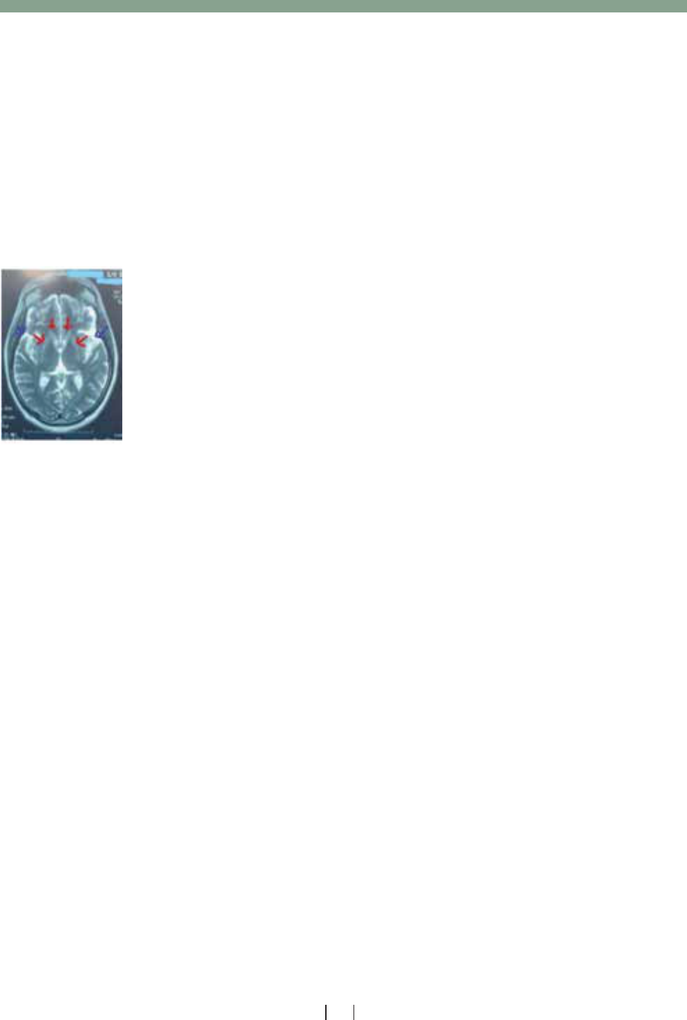
urinary copper 48 ug/day (<60 ug/day), SGPT 32U/L (<40 U/L). ANA was positive. Magnetic resonance imaging (MRI)
brain showed prominence of the cortical sulci and ventricles representing brain involution not corresponding to
patient’s age (figure 1) .Patient was on thyroxin 100 ug per day , levodopa/carbidopa 25/250 mg half tablet thrice a
day (TDS) and trihexiphenidyl 2 mg half TDS titrated upto 2 mg TDS ,Baclofen 10 mg half BD titrated gradually to 1
TDS along with rehabilitation . After a follow up of 4 weeks, his oromandibular dystonia improved to some extent but
no significant improvement in his leg dystonia and bradykinesia .Whether patient should be started on glutein free diet
as he was asymptomatic for coeliac disease and immunotherapy on the basis of work up or not to improve patient’s
dystonia was still undecided. According to the available literature patient was planned for immunotherapy with regular
follow up of his symptoms for the response.
FIGURE 1: MRI T2W image of case 2 showing prominence of the cortical sulci and
ventricles representing brain involution not corresponding to patient’s age (Blue arrows)
.Hyper intense signal are seen in bilateral basal ganglia (Red arrows).
DISCUSSION
Autoimmune movement disorders in neurology are emerging and likewise the associated
autoantibodies which need to be worked up. A number of essential tests as the supportive
evidence are recommended to be performed in case of suspected autoimmune movement
disorder. Antibody-related dystonia differs from primary dystonia, as it follows a characteristic
pattern in body parts affected and age at onset. In a patient with adult onset dystonia,
secondary causes of dystonia are more common. Work up is needed for the secondary causes
which turned out to be autoimmune in origin as our patient had significant abnormalities in work up which suggest a
systemic autoimmune involvement. Antibody associated dystonia typically presents as a combined dystonia means in
combination with further clinical signs, in particular encephalopathy
9
Neurodegeneration as evident on MRI brain of
my case 2 patient with OMD and limb dystonia is supporting this feature. Moreover he was asymptomatic for celiac
disease and thyroid disorder and surprisingly had very high CPK without any evidence of myopathy.
American academy of neurology published a small study in 2019 comprising of 4 patients diagnosed with autoimmune
dystonia. Generalized asymmetric dystonia was the most common presentation, and parkinsonian symptoms (rigidity
and resting tremors) were present in 1 patient. serum and cerebrospinal autoimmune encephalitis work up were
inconclusive. All patients were treated with Immunotherapy using intravenous methylprednisolone 1 g daily for 5 days
and intravenous immunoglobulin 0.4 gm/kg for 5 days, followed by azathioprine maintenance dose (2 mg/kg).
Improvement was found in all cases with complete resolution of autoimmune dystonia in 2 cases. Hence, it was
concluded that despite a significant delay in therapy due to misdiagnosis, autoimmune dystonia patients may respond
well to immunomodulators with an encouraging outcome
10
. Among the disorders associated with autoimmune disease
, Celiac disease (CD) is emerging as being frequently reported with significantly increased prevalence in individuals
with CD and their first-degree relatives as compared to controls
11-13
.The estimated burden of Autoimmune disease
(AD) in CD cases up to 15%
10
.It has a wide clinical spectrum which includes specific features of malabsorption with
diarrhoea, non-specific extra intestinal features, subclinical or asymptomatic variants , and likely disease
characterized by positive serology with a normal biopsy of intestinal mucosa
14,15
. The risk factors for developing other
AD are an early diagnosis and a positive family history of autoimmunity, whereas the gluten-free diet (GFD) has a
beneficial role
16
. Another article concluded that autoimmune movement disorders with an extracellular antibody and
without cancer are more likely to respond to treatment while the disorders with a cancer and/or an intracellular
antibody respond less well to immunotherapy
2
.Furthermore, while a GFD is found to improve and reverse symptoms
of gluten-associated ataxia and is an established therapy for CD, it is not the case with oromandibular dystonia (OMD).
CD has been found increasingly in patients with autoimmune thyroid disease i.e. Grave’s disease and Hashimoto’s
thyroiditis ,having a 2% to 7% prevalence
5
.Several mechanisms are suggested about the co-occurrence of CD and
autoimmune thyroid disease like similar genetic predisposition and both having the gene encoding cytotoxic
T-lymphocyte-associated antigen .In addition, tTG2 IgA antibodies has been shown to react with thyroid tissue, leading
to the development of thyroid disease in CD.In our both cases patients had positive work up for celiac disease as well
as had evidence of thyroid dysfunction along with the movement disorder.

References:
1.(https://pathology.jhu.edu/autoimmune/prevalence)
( john hopkin’s medicine)
2. Conor Fearon, Orna O’Toole.Autoimmune Movement
Disorders. Semin Neurol 2018; 38:316–329.
3. Honorat JA, McKeon A: Autoimmune movement
disorders: a clinical and laboratory approach. Curr
Neurol Neurosci Rep. 2017;17(1):4. doi:
10.1007/s11910-017-0709-2.
4. Jones AL, Flanagan EP, Pittock SJ, et al. Responses
to and outcomes of treatment of autoimmune
cerebellar ataxia in adults. JAMA Neurol.
2015;72(11):1304–1312. doi:10.1001/
jamaneurol.2015.2378.)
5. Lauret E, Rodrigo .Celiac Disease and
Autoimmune-Associated Conditions. BioMed Research
International Volume2013,Article ID 127589, 17
pages http://dx.doi.org/10.1155/2013/127589.
6. https://doi.org/10.14802/jmd.16063 / J Mov
Disord 2017;10(2):105-107 pISSN 2005-940X /
eISSN 2093-493)
7.Donaldson I, Marsden CD,Schneider S. Marsden’s
Book of Movement Disorders. Oxford: Oxford University
Press; 2012.
8 . Baizabal-Carvallo JF, Jankovic J .Autoimmune
and paraneoplastic movement disorders: An
update. REVIEW ARTICLE| VOLUME 385,
P175-184, FEBRUARY 15, 2018.
DOI:https://doi.org/10.1016/j.jns.2017.12.035
9 . Gövert F, Leypoldt F , Wandinger KP et al.
Antibody-related movement disorders – a
comprehensive review of phenotype autoantibody
correlations and a guide to testing Neurological
Research and Practice (2020) 2:6
https://doi.org/10.1186/s42466-020-0053-x
10.American academy of neurology ( April 09, 2019;
92 (15 Supplement) MAY 6, 2019) .
11.F.Cataldo and V.Marino, “Increased prevalence of
autoimmune diseases in first-degree relatives of
patients with celiac disease,” Journal of Pediatric
Gastroenterology and Nutrition,vol.36,no.
4,pp.470–473,2003.
12. M. Viljamaa, K. Kaukinen, H. Huhtala, S.
Kyr¨onpalo, M. Rasmussen, and P. Collin, “Coeliac
disease, autoimmune diseases and gluten
exposure,”Scandinavian Journal of Gastroenterology,
vol.40,no.4,pp.437–443,2005.
13. S. L. Neuhausen, L. Steele, S. Ryan et al.,
“Co-occurrence of celiac disease and other
autoimmune diseases in celiacs and their first-degree
relatives,” Journal of Autoimmunity,vol.31, no.
2,pp.160–165,2008.
14. P. Collin, T. Reunala, E. Pukkala, P. Laippala, O.
Keyril¨ainen, and A. Pasternack, “Coeliac
disease—associated disorders and
survival,”Gut,vol.35,no.9,pp.1215–1218,1994.
15. A. Fasano, I. Berti, T. Gerarduzzi et al.,
“Prevalence of Celiac disease in at-risk and
not-at-risk groups in the United States : a large
multicentre study, ”Archives of Internal
Medicine,vol.163, no.3,pp.286–292,2003.
16 .J. Cosnes, C. Cellier, S. Viola et al., “Incidence of
autoimmune diseases in celiac disease: protective
effect of the gluten-free diet,” Clinical
Gastroenterology and Hepatology,vol.6,no.7,pp.
753–758,2008.
PAKISTAN JOURNAL OF NEUROLOGICAL SCIENCES
33
VOL. 15 (2) APRIL-JUNE 2020
In 2017, “Isolated Neurological Manifestation in Silent Celiac Disease” was published in which a case of idiopathic
oromandibular dystonia (OMD) is discussed that was unresponsive to traditional symptomatic therapy. However,
complete resolution of symptoms was obtained after celiac disease (CD) diagnosis and gluten free diet
6
. In the same
way, our case 1 patient also showed the improvement. Association of asymptomatic high creatine phosphokinase
(CPK) level and celiac disease or other neurological illnesses remains unexplained except for the limited clue due to
its types CK MM, CK BB, and CK MB. Further studies are highly recommended to clarify the unexplained factors and
further management of such cases.
CONCLUSION:
Isolated autoimmune movement disorders are uncommon and are usually associated with other neurological features.
A prompt and thorough work up for an underlying cause and associated neural or systemic antibodies in such
suspected cases. Ultimately through this approach decisions can be easily taken for the treatment and prognosis of
disease. Further studies are direly needed to clarify the issues encountered in management of such cases.

References:
1.(https://pathology.jhu.edu/autoimmune/prevalence)
( john hopkin’s medicine)
2. Conor Fearon, Orna O’Toole.Autoimmune Movement
Disorders. Semin Neurol 2018; 38:316–329.
3. Honorat JA, McKeon A: Autoimmune movement
disorders: a clinical and laboratory approach. Curr
Neurol Neurosci Rep. 2017;17(1):4. doi:
10.1007/s11910-017-0709-2.
4. Jones AL, Flanagan EP, Pittock SJ, et al. Responses
to and outcomes of treatment of autoimmune
cerebellar ataxia in adults. JAMA Neurol.
2015;72(11):1304–1312. doi:10.1001/
jamaneurol.2015.2378.)
5. Lauret E, Rodrigo .Celiac Disease and
Autoimmune-Associated Conditions. BioMed Research
International Volume2013,Article ID 127589, 17
pages http://dx.doi.org/10.1155/2013/127589.
6. https://doi.org/10.14802/jmd.16063 / J Mov
Disord 2017;10(2):105-107 pISSN 2005-940X /
eISSN 2093-493)
7.Donaldson I, Marsden CD,Schneider S. Marsden’s
Book of Movement Disorders. Oxford: Oxford University
Press; 2012.
8 . Baizabal-Carvallo JF, Jankovic J .Autoimmune
and paraneoplastic movement disorders: An
update. REVIEW ARTICLE| VOLUME 385,
P175-184, FEBRUARY 15, 2018.
DOI:https://doi.org/10.1016/j.jns.2017.12.035
9 . Gövert F, Leypoldt F , Wandinger KP et al.
Antibody-related movement disorders – a
comprehensive review of phenotype autoantibody
correlations and a guide to testing Neurological
Research and Practice (2020) 2:6
https://doi.org/10.1186/s42466-020-0053-x
10.American academy of neurology ( April 09, 2019;
92 (15 Supplement) MAY 6, 2019) .
11.F.Cataldo and V.Marino, “Increased prevalence of
autoimmune diseases in first-degree relatives of
patients with celiac disease,” Journal of Pediatric
Gastroenterology and Nutrition,vol.36,no.
4,pp.470–473,2003.
12. M. Viljamaa, K. Kaukinen, H. Huhtala, S.
Kyr¨onpalo, M. Rasmussen, and P. Collin, “Coeliac
disease, autoimmune diseases and gluten
exposure,”Scandinavian Journal of Gastroenterology,
vol.40,no.4,pp.437–443,2005.
13. S. L. Neuhausen, L. Steele, S. Ryan et al.,
“Co-occurrence of celiac disease and other
autoimmune diseases in celiacs and their first-degree
relatives,” Journal of Autoimmunity,vol.31, no.
2,pp.160–165,2008.
14. P. Collin, T. Reunala, E. Pukkala, P. Laippala, O.
Keyril¨ainen, and A. Pasternack, “Coeliac
disease—associated disorders and
survival,”Gut,vol.35,no.9,pp.1215–1218,1994.
15. A. Fasano, I. Berti, T. Gerarduzzi et al.,
“Prevalence of Celiac disease in at-risk and
not-at-risk groups in the United States : a large
multicentre study, ”Archives of Internal
Medicine,vol.163, no.3,pp.286–292,2003.
16 .J. Cosnes, C. Cellier, S. Viola et al., “Incidence of
autoimmune diseases in celiac disease: protective
effect of the gluten-free diet,” Clinical
Gastroenterology and Hepatology,vol.6,no.7,pp.
753–758,2008.
Conflict of interest: There is no conflict of interest..
Funding disclosure: Nil
Author’s contribution:
Ramla Nayaib Hashmi; data collection, data analysis, manuscript writing, manuscript review
Ummul Kiram; data analysis,manuscript writing, manuscript review
Rabbia Tariq; data analysis, manuscript writing, manuscript review
PAKISTAN JOURNAL OF NEUROLOGICAL SCIENCES
34
VOL. 15 (2) APRIL-JUNE 2020
