
Vol. 3, No. 3 Modern Applied Science
124
Experimental Investigation on the Effects of Audible Sound
to the Growth of Escherichia coli
Joanna Cho Lee Ying
School of Science and Technology, Universiti Malaysia Sabah
Lock Bag 2073, 88999 Kota Kinabalu, Sabah, Malaysia
E-mail: joanna_25[email protected]om
Jedol Dayou
Vibration and Sound Research Group (VIBS), Universiti Malaysia Sabah
Lock Bag 2073, 88999 Kota Kinabalu, Sabah, Malaysia
Tel: 60-88-320-302 E-mail:
Chong Khim Phin (Corresponding author)
School of Sustainable Agriculture, Universiti Malaysia Sabah
Lock Bag 2073, 88999 Kota Kinabalu, Sabah, Malaysia
Tel: 60-88-325-655 E-mail:
Abstract
In this paper, we report an experimental result regarding the effects of audible sound on the growth of Escherichia coli
(
E. coli). Standardized E. coli suspensions of fixed concentration were used for inoculation throughout the experiment
in nutrient agar (NA) and nutrient broth (NB). First, the samples were incubated at 37ºC for three hours in a water
bath-shaker for NB and in a conventional oven for NA. The samples were then transferred to an acoustic chamber
JedMark LV-1 with given sound treatment at controlled temperature of 24±2ºC for five hours for NB and 16 hours for
NA. Three different tonal frequencies were selected for sound treatment in this experiment which is 1 kHz, 5 kHz and
15 kHz. The growth of E. coli was assessed by their cell number through indirect viable cell counts (
E. coli on NA) and
direct viable cell counts (
E. coli on NB), after the incubation with sound in the acoustic chamber. We found that all
selected frequencies were able to promote the growth of
E. coli. In particular, the tonal sound of 5 kHz gave significant
increase in cell number of
E. coli for both growth media.
Keywords: Escherichia coli, Audible sound wave, Plate count, Direct microscopic count
1. Introduction
Mechanical waves have been shown to have effect on microbes. Ultrasound for example, has been used for sterilizing
and killing unwanted bacteria due to thinning of cell membranes, localized heating and production of free radicals
(Piyasena et al., 2003). Ultrasound is able to inactivate bacteria and deagglomerate bacterial clusters or flocks through a
number of physical, mechanical and chemical effects arising from acoustic cavitations (Joyce et al., 2003). However,
certain frequency of ultrasound was found to increase the growth rate of bacteria cells such as
E. coli, Staphylococcus
epidermidis
and Pseudomonas aeruginosa cells adhered to and grew on a polyethylene surface (Pitt and Ross, 2003).
Ultrasound technology is relatively expensive to be used for large scale microbiological decontamination or production.
The consideration of the amount of
energy inputs and its cost must be first carried out if this technology is put to be
used
(Hao et al., 2004). A more affordable alternative way using audible sound wave may sound practical, and there
have been some evidences for this possibility. Matsuhashi
et al. (1998) for example, had found that audible sound and
ultrasound waves between 6 kHz to 40 kHz can
induce colony formation of Bacillus carbophilus grown on
non-permissive media.
In this paper, the effect of audible sound of certain frequencies to E. coli is investigated as a
compliment to the application of ultrasound. The effect is assessed
based on the comparison in their cell number,
through direct and indirect viable cell
counts, with their respective control samples.

Modern Applied Science March, 2009
125
2. Materials and methods
2.1 Esherichia coli
The investigation on the effects of audible sound to E. coli was carried out in two modes - on the E. coli that was
inoculated on NA and on the
E. coli that was inoculated on NB medium. The E. coli on NA was used for indirect viable
cell count using plate counts whereas the
E. coli on NB was used for direct viable cell counts using a haemocytometer.
E. coli was obtained from previous stock cultured at Microbiology Lab, Universiti Malaysia Sabah and maintained on
NA plate (medium) at 37ºC for 24 hours in an oven. The medium was prepared by suspending 10g of NA in 500ml
distilled water and was autoclaved to sterilize at 121ºC for two hours. This is the secondary cultured
E. coli to be used
to prepare samples in the experiment.
2.2 Innoculation
Eight NB and six NA media were prepared in this experiment. The NB medium was prepared by suspending 4g of NB
in 500ml distilled water and autoclaved to sterilize at 121ºC for two hours whereas NA medium was prepared in the
same way described previously. Single colony of
E. coli from the secondary culture was transferred to each NB
mediums which was prepared in screwed-capped bottles. The remainder of the secondary cultured
E. coli was diluted
ten-folds and then divided into eight for inoculation on NA which was prepared on petri dishes.
2.3 Incubation of the samples
The NB samples were incubated in water bath-shaker at 100 rotations per minute at 37
o
C for 3 hours and the NA
samples with
E. coli were incubated in conventional oven also at 37
o
C for 3 hours. After the incubation, the samples
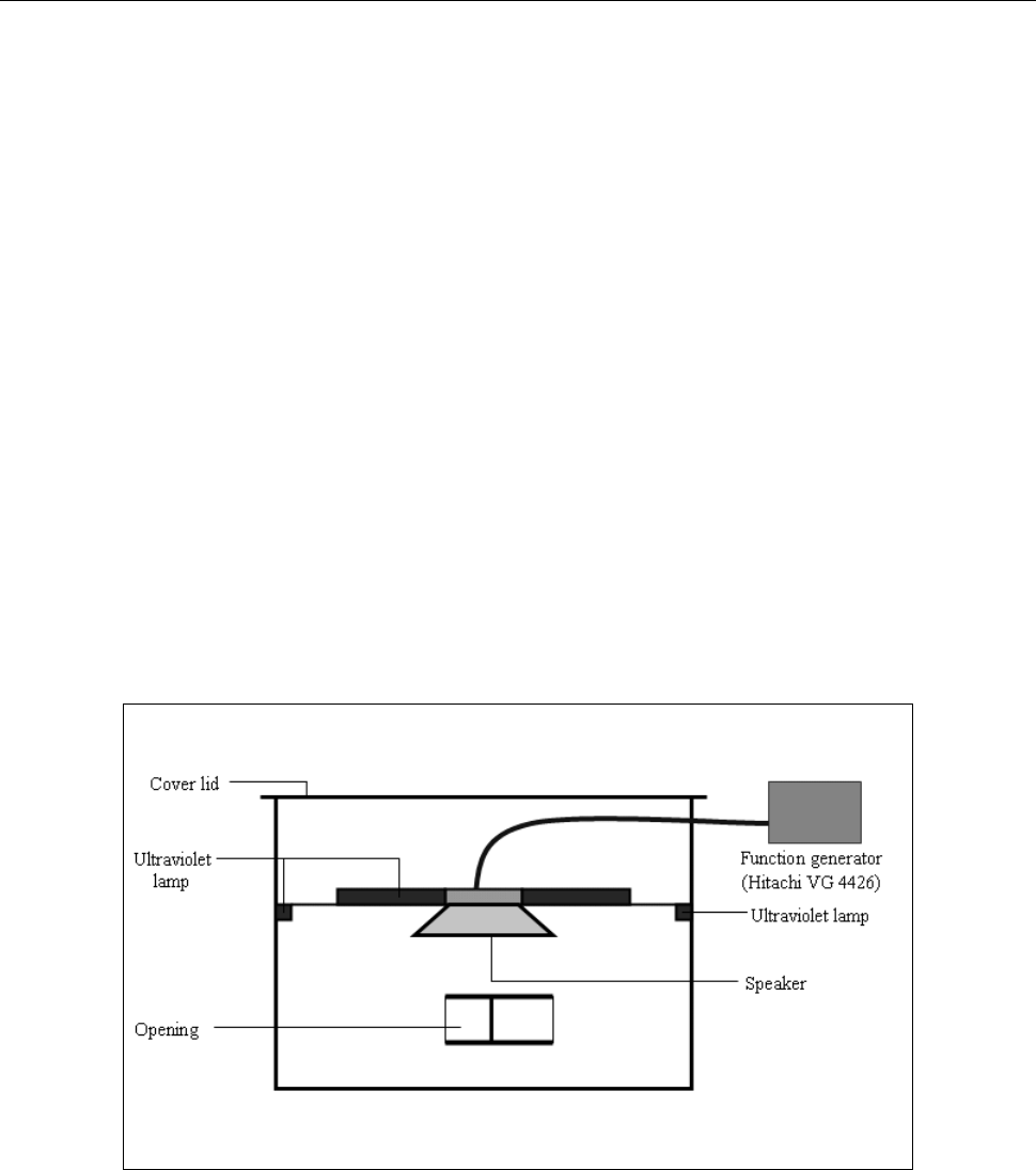
were then transferred to an acoustic chamber JedMark LV-1 (Figure 1) for sound treatment through the opening. The
chamber which is made of thick glass, is air tight when in operation and equipped with three ultraviolet lamps for fully
sterilizing the inner part of the chamber before the treatment carried out. The chamber was also installed with a
loudspeaker connected to a function generator so that tonal frequency of sound can be generated. The samples were
exposed to sound for five hours at frequency of 1 kHz, 5 kHz and 15 kHz for sound treatment. For each selected
frequency, two NA and NB samples were exposed simultaneously whereas two control samples was maintained without
sound treatment as reference as reference. During the treatment process, the temperature was maintained at 24±2ºC.
2.4 Measurements
Soon after the exposure, direct E. coli cell counts was performed on NB samples using haemocytometer whereas
indirect cell counts was carried out on NA samples using plates counts technique. The same
E. coli cell counting
method was performed on the respective control medium type. In the direct cell counts, trypan blue (0.4% w/v) was
used to stain dead cells (stained dark blue and immobile). In indirect cell counts, the number of viable colonies growth
on NA plates was counted with the assumption that the number of
E. coli cells is proportional to the number of colonies
formed on the media which is measured in colony forming unit per milliter, CFU.
3. Results and discussion
3.1 Direct viable cell count
As previously mentioned, direct viable cell counts method was used to determine the number of viable E. coli cell on
the NB samples using a hemocytometer. Throughout the experiment, less than one percent of dead cells were found in
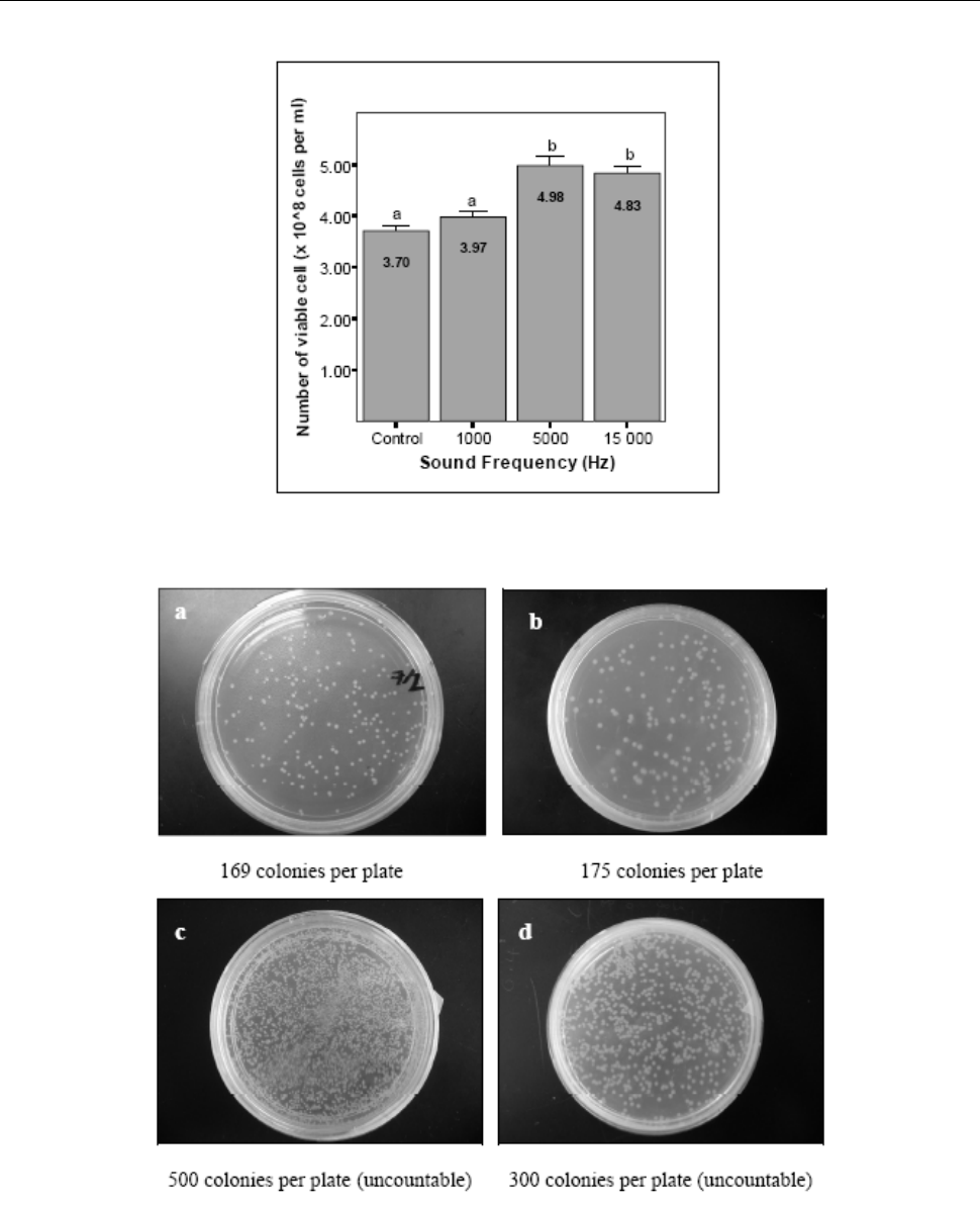
each NB samples. It was found that sound treatment at all selected frequencies have increased the number of viable
cells compared to the control samples. Sound treatment at 1 kHz for example was found to increase the average number
of viable cells to 3.97 x 10
8
cells per ml, 4.98 x 10
8
cells per ml for the sound at 5 kHz and 4.83 x 10
8
cells per ml for
the sound at 15 kHz. These increases are equivalent to 7%, 34% and 30.5% for the sound treatment at 1 kHz, 5 kHz and
15 kHz, respectively, compared to the average number of viable cells found on the control sample which is only 3.70 x
10
8
cells per ml. The cell number for this direct viable cell counts is given in Figure 2. From the observations, sound
treatment at 5 kHz found to give the most significant increases in the number of viable cells followed by the sound at 15
kHz and 1 kHz.
3.2 Indirect viable cell count
Indirect viable cell counts (or plate counts) was performed to determine the viable E. coli cells on NB samples using
ordinary microscope. The assumption is each
E. coli cell give rise to one colony, and therefore the indirect viable cell
count is measured in colony forming unit per milliter, CFU. As in the direct viable cell count, it was found that sound
treatment at all selected frequencies also have increased the CFU of
E. coli (and so the number of E. coli cells). Sound
treatment at 1 kHz for example increased the average CFU to 1.75 x 10
9
compared to only 1.69 x 10
9
CFU for the
control sample. On the other hand, sound treatment at 5 kHz and 15 kHz gave rise to uncountable colony. However,
visual inspection shows that sound treatment at 5 kHz gave higher value of CFU compared to frequency 15 kHz. The
resultant increased for the sound treatment at 1 kHz is 3.5% compared to the control samples. The mean of
E. coli CFU
is shown in Table 1 whereas Figure 3 represents the visual inspection of the samples. In Table 1 and Figure 3, the mean
Vol. 3, No. 3 Modern Applied Science
126
number of CFU for 5 kHz and 15 kHz sound treatments were simply given so that they can be distinguished.
4. Conclusion
In this paper, we presented the result of an experimental investigation on the effects of audible sound to the growth of E.
coli
inoculated on NA and NB media. We found that all selected frequencies (1 kHz, 5 kHz and 15 kHz) have increased
the number of viable cells of the bacteria. This shows that the bacteria react positively to the given sound treatment
which results in the growth of the number of the bacteria cells. However, the degree of their respond is different to a
different sound frequency. This is consistence with the finding reported by other researchers. In this experiment, we
found that
E. coli on NA and NB responded more to sound at frequency 5 kHz compared to other frequencies. This
shows that
E. coli has selective frequency response towards sound treatment.
References
Hao, H., Wu, M., Chen, Y., Tang, J. & Wu, Q. (2004) Cavitations mechanism in cyanobacterial growth inhibition by
ultrasonic irradiation.
Colloids and Surfaces, 33(3-4):151-156.
Joyce, E., Phull, S.S., Lorimer, J.P. & Mason, T.J. (2003). The development and evaluation of ultrasound for the
treatment of bacterial suspensions. A study of frequency, power and sonification time on cultured bacillus species.
Ultrasonics Sonochemistry, 10(6):315-318.
Matsuhashi, M., Pankrushina, A.N., Takeuchi, S., Ohshima, H., Miyoi, H., Endoh, K., Murayama, K., Watanabe, H.,
Endo, S., Tobi, M., Mano, Y., Hyodo, M., Kobayashi, T., Kaneko, T., Otani, S., Yoshimura, S., Harata, A. & Sawada, T.
(1998). Production of sound waves by bacterial cells and the response of bacterial cells to sound.
Journal of General
Applied Microbiology
, 44: 49-55.
Pitt, W.G. & Ross, S.A. (2003).Ultrasound Increases the Rate of Bacterial Cell Growth.
Biotechnology Progress,
19(3):1038-1044.
Piyasena, P., Mohareb, E. & McKellar, R.C. (2003). Inactivation of microbes using ultrasound: A review
. International
Journal of Food Microbiology
, 87(3):207-216.
Figure1. Schematic diagram of the JedMark LV-1 acoustic chamber
Modern Applied Science March, 2009
127
Figure 2. Viable
E. coli cell count (x10
8
cells per ml) for different sound treatments
Figure 3. Number of colony per plate for the different sound treatments.
(a) Control, (b) 1kHZ, (c) 5 kHZ and (d) 15kHZ
